Taryn Roton
  • Blog
  • About
  • Contact

The Best Ways To Spot Calcaneal Spur

9/22/2015

 
Inferior Calcaneal Spur

Overview

Heel spur is a hook of bone that forms on the heel bone. The condition itself is not the most painful; it is the inflammation and irritation that cause the heel pain. Often times, plantar fasciitis is a cause of heel spurs. When the ligaments are pulled away, calcium deposits form on the hooked bone. An orthotic will help relieve the pain associated with heel spurs.

Causes

Some causes of heel spurs include abnormal or lopsided walking, which places excessive stress on the heel bone, ligaments and nerves Running, jogging or jumping, especially on hard surfaces. Poorly fitted or badly worn shoes, especially those lacking appropriate arch support, excess weight and obesity.

Calcaneal Spur

Symptoms

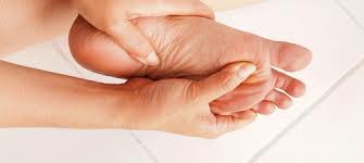
Heel spurs may or may not cause symptoms. Symptoms are usually related to the plantar fasciitis. You may experience significant pain. Your heel pain may be worse in the morning when you first wake up or during certain activities.

Diagnosis

Diagnosis of a heel spur can be done with an x-ray, which will be able to reveal the bony spur. Normally, it occurs where the plantar fascia connects to the heel bone. When the plantar fascia ligament is pulled excessively it begins to pull away from the heel bone. When this excessive pulling occurs, it causes the body to respond by depositing calcium in the injured area, resulting in the formation of the bone spur. The Plantar fascia ligament is a fibrous band of connective tissue running between the heel bone and the ball of the foot. This structure maintains the arch of the foot and distributes weight along the foot as we walk. However, due to the stress that this ligament must endure, it can easily become damaged which commonly occurs along with heel spurs.

Non Surgical Treatment

If pain and other symptoms of inflammation-redness, swelling, heat-persist, you should limit normal daily activities and contact a doctor of podiatric medicine. The podiatric physician will examine the area and may perform diagnostic X-rays to rule out problems of the bone. Early treatment might involve oral or injectable anti-inflammatory medication, exercise and shoe recommendations, taping or strapping, or use of shoe inserts or orthotic devices. Taping or strapping supports the foot, placing stressed muscles and tendons in a physiologically restful state. Physical therapy may be used in conjunction with such treatments. A functional orthotic device may be prescribed for correcting biomechanical imbalance, controlling excessive pronation, and supporting of the ligaments and tendons attaching to the heel bone. It will effectively treat the majority of heel and arch pain without the need for surgery. Only a relatively few cases of heel pain require more advanced treatments or surgery. If surgery is necessary, it may involve the release of the plantar fascia, removal of a spur, removal of a bursa, or removal of a neuroma or other soft-tissue growth.

Surgical Treatment

In a small number of cases (usually less than 5 percent), patients may not experience relief after trying the recommendations listed above. It is important that conservative treatments (such as those listed above) be performed for AT LEAST a year before considering surgery. Time is important in curing the pain from heel spurs, and insufficient treatment before surgery may subject you to potential complications from the procedure. If these treatments fail, your doctor may consider an operation to loosen the plantar fascia, called a plantar fascia release.

Chronic Bursitis Of The Foot

8/24/2015

 
Overview

This is a very common condition that leads to foot pain . Once you understand what it actually is you will wonder why it does not occur more often, particularly in the foot. It is an inflamed bursal sac. A bursal sac is a sac filled with fluid that acts to lubricate and reduce the friction between two surfaces in the body, usually muscles and tendons as they glide over bony prominences, however their purpose in not limited to just muscles and tendons. The body contains literally hundreds of bursal sacs but in the foot there is just one naturally occurring (adventitious) bursal sac. It is located between the Achilles tendon and the heel bone (calcaneaus), otherwise known as an achilles tendon bursal sac.In this instance the Achilles tendon is protected from the pressure of the heel bone pressing against it when we walk.

Causes

A rapid increase in physical activity levels or thinning of the heel?s protective fat pad are factors that may contribute to infracalcaneal bursitis. Other possible causes of infracalcaneal bursitis include blunt force trauma, acute or chronic infection, and arthritic conditions. The following factors may increase your risk of experiencing bursitis, including infracalcaneal bursitis. Participating in contact sports. Having a previous history of bursitis in any joint. Poor conditioning. Exposure to cold weather. Heel striking when running, especially in conventional running shoes with heel elevation.

Symptoms

Some of the symptoms of bursitis in the heel, or retrocalcaneal bursitis, are as described below. Severe pain in the heel area of the foot, sometimes radiating to the ankle, associated with physical activities like walking, jogging and even on physical contact to the area. The physical signs of heel bursitis, which are noticeable in the heel area, are reddish discoloration of the skin that is warm to touch.

Diagnosis

A good clinical practise includes evaluation of the tendon, bursa and calcaneum by, careful history, inspection of the region for bony prominence and local swelling as well as palpation of the area of maximal tenderness. Biomechanical abnormalities, joint stiffness and proximal soft tissue tightening can exacerbate an anatomical predisposition to retrocalcaneal bursitis, they warrant correction when present.

Non Surgical Treatment

Caregivers may give you special shoe inserts with a cutout around the tender area. You may also be told to wear shoes with a reinforced heel counter. This will give better heel control. You may need other shoe inserts (wedges) to raise your heel so it does not press against the back of the shoe. You may also wear shoes that are open in the back, such as sandals that have no strap across the heel. You may use ibuprofen (eye-bu-PROH-fen) and acetaminophen (a-seet-a-MIN-oh-fen) medicine for your pain. These may be bought over-the-counter at drug or grocery stores. Do not take ibuprofen if you are allergic to aspirin. You may be given shots of medicine called steroids (STER-oids) to decrease inflammation. Caregivers may add local anesthesia (an-es-THEE-zah) to the steroids. This medicine helps decrease bursitis pain. Because these shots decrease swelling and pain, you may feel like your ankle is healed and that you can return to heavy exercise. It is important to not exercise until your caregiver says it is OK. You could make the bursitis worse if you exercise too soon. You may need surgery to remove the bursa or part of your ankle bone. Surgery is usually not necessary unless the bursitis is very bad and does not heal with other treatments. Your caregiver may want you to go to physical (FIZ-i-kal) therapy (THER-ah-pee). Physical therapists may use ultrasound to increase blood flow to the injured area. Caregivers may use massage to stretch the tissue and bring heat to the injury to increase blood flow. These and other treatments may help the bursitis heal faster. Exercises to stretch your Achilles tendon and make it stronger will be started after the bursitis has healed. You may gradually increase the amount of weight you put on your foot when caregivers say it is OK. You may be told to stop exercising if you feel any pain.

Surgical Treatment

Surgery. Though rare, particularly challenging cases of retrocalcaneal bursitis might warrant a bursectomy, in which the troublesome bursa is removed from the back of the ankle. Surgery can be effective, but operating on this boney area can cause complications, such as trouble with skin healing at the incision site. In addition to removing the bursa, a doctor may use the surgery to treat another condition associated with the retrocalcaneal bursitis. For example, a surgeon may remove a sliver of bone from the back of the heel to alter foot mechanics and reduce future friction. Any bone spurs located where the Achilles attaches to the heel may also be removed. Regardless of the conservative treatment that is provided, it is important to wait until all pain and swelling around the back of the heel is gone before resuming activities. This may take several weeks. Once symptoms are gone, a patient may make a gradual return to his or her activity level before their bursitis symptoms began. Returning to activities that cause friction or stress on the bursa before it is healed will likely cause bursitis symptoms to flare up again.

Prevention

Maintain proper form when exercising, as well as good flexibility and strength around the ankle to help prevent this condition. Proper stretching of the Achilles tendon helps prevent injury.

Recuperating From Bunion And Hammertoe Surgery

7/5/2015

 
HammertoeOverview

hammertoes is a contracture (bending) of one or both joints of the second, third, fourth, or fifth (little) toes. This abnormal bending can put pressure on the toe when wearing shoes, causing problems to develop. Hammertoes usually start out as mild deformities and get progressively worse over time. In the earlier stages, hammer toes are flexible and the symptoms can often be managed with noninvasive measures. But if left untreated, hammer toes can become more rigid and will not respond to non-surgical treatment. Because of the progressive nature of hammertoes, they should receive early attention. Hammertoes never get better without some kind of intervention.

Causes

Risk factors for Hammer toes hammertoe include heredity, a second toe that is longer than the first (Morton foot), high arches or flat feet, injury in which the toe was jammed, rheumatoid arthritis, and, in diabetics, abnormal foot mechanics resulting from muscle and nerve damage. Hammertoe may be precipitated by advancing age, weakness of small muscles in the foot (foot intrinsic muscles), and the wearing of shoes that crowd the toes (too tight, too short, or with heels that are too high). The condition is more common in females than in males.

Hammer ToeSymptoms

A toe stuck in an upside-down "V" is probably a hammertoe. Some symptoms are, pain at the top of the bent toe when putting on a shoe. Corns forming on the top of the toe joint. The toe joint swelling and taking on an angry red colour. Difficulty in moving the toe joint and pain when you try to so. Pain on the ball of the foot under the bent toe. Seek medical advice if your feet regularly hurt, you should see a doctor or podiatrist. If you have a hammertoe, you probably need medical attention. Ask your doctor for a referral to a podiatrist or foot surgeon. Act now, before the problem gets worse.

Diagnosis

Some questions your doctor may ask of you include, when did you first begin having foot problems? How much pain are your feet or toes causing you? Where is the pain located? What, if anything, seems to improve your symptoms? What, if anything, appears to worsen your symptoms? What kind of shoes do you normally wear? Your doctor can diagnose hammertoe or mallet toe by examining your foot. Your doctor may also order X-rays to further evaluate the bones and joints of your feet and toes.

Non Surgical Treatment

Any forefoot problems that cause pain or discomfort should be given prompt attention. Ignoring the symptoms can aggravate the condition and lead to a breakdown of tissue, or possibly even infection. Conservative treatment of mallet toes begins with accommodating the deformity. The goal is to relieve pressure, reduce friction, and transfer forces from the sensitive areas. Shoes with a high and broad toe box (toe area) are recommended for people suffering from forefoot deformities such as mallet toes. This prevents further irritation in the toe area from developing. Other conservative treatment includes forefoot supports such as gel toe caps, gel toe shields and toe crests. Gel forefoot supports provide immediate comfort and relief from common forefoot disorders without drying the skin.

Surgical Treatment

If this fails or if treatment is not sought until the toes are permanently misaligned, then surgery may be required. Surgery may involve either cutting the tendon or fusing the joint. Congenital conditions should be treated in early childhood with manipulations and splinting.

Hammer Toe Treatments Without Surgery

7/1/2015

 
Hammer ToeOverview

Hammer toe is a secondary problem originating from fallen cross arches. The toes start to curl and get pulled backwards, as the collapsed or pushed out metatarsal bones pull the tendons and ligaments, and causes them to get shorter and tighter. This condition causes the toes have higher pressure and they have limited movement and cannot be straightened fully. This can lead to numbness and pain in the toes as muscles, nerves, joints and little ligaments are involved with this condition. As the top part of the toe can rub against the shoe, it can cause corns and calluses.

Causes

Hammertoes are most common in women, and a big part of this is poor shoe choices, which are a big factor in the development of many foot problems. Tight toe boxes and high heels are the biggest culprits. Genetics certainly plays a role in some cases of hammertoes, as does trauma, infection, arthritis, and certain neurological and muscle disorders. But most cases of contracted toes are associated with various biomechanical abnormalities in how a patient walks. This causes the muscles and tendons to be used excessively or improperly, which deforms the toes over time.

HammertoeSymptoms

A toe stuck in an upside-down "V" is probably a hammertoe. Some symptoms are, pain at the top of the bent toe when putting on a shoe. Corns forming on the top of the toe joint. The toe joint swelling and taking on an angry red colour. Difficulty in moving the toe joint and pain when you try to so. Pain on the ball of the foot under the bent toe. Seek medical advice if your feet regularly hurt, you should see a doctor or podiatrist. If you have a hammertoe, you probably need medical attention. Ask your doctor hammertoe for a referral to a podiatrist or foot surgeon. Act now, before the problem gets worse.

Diagnosis

Your healthcare provider will examine your foot, checking for redness, swelling, corns, and calluses. Your provider will also measure the flexibility of your toes and test how much feeling you have in your toes. You may have blood tests to check for arthritis, diabetes, and infection.

Non Surgical Treatment

People with a hammer toe benefit from wearing shoes in which the toe box is made of a flexible material and is wide enough and high enough to provide adequate room for the toes. High-heeled shoes should be avoided, because they tend to force the toes into a narrow, flat toe box. A doctor may recommend an insert (orthotic) for the shoe to help reduce friction and pressure on the hammer toe. Wearing properly fitted shoes may reduce pain and inflammation. It may also prevent ulcers from developing and help existing ulcers heal. However, the hammer toe does not disappear.

Surgical Treatment

Joint resection procedures involves removing part of one of the two small joints of the toe directly underneath where the digit is crooked. The purpose is to make room for the toe to be re-positioned flat or straight. Because hammer toes become rigid or fixed with time, removing the joint becomes the only option when the knuckle is stiff. Its important to understand that this procedure does not involve the joint of the ball of the foot, rather the a small joint of the toe. Medical terminology for this procedure is called a proximal interphalangeal joint arthroplasty or a distal interphalangeal joint arthroplasty, with the latter involving the joint closer to the tip of the toe.

Hammer ToePrevention

Early Development. The first year of life is important for foot development. Parents should cover their babies' feet loosely, allowing plenty of opportunity for kicking and exercise. Change the child's position frequently. Children generally start to walk at 10 - 18 months. They should not be forced to start walking early. Wearing just socks or going barefoot indoors helps the foot develop normally and strongly and allows the toes to grasp. Going barefoot outside, however, increases the risk for injury and other conditions, such as plantar warts. Children should wear shoes that are light and flexible, and since their feet tend to perspire, their shoes should be made of materials that breathe. Replace footwear every few months as the child's feet grow. Footwear should never be handed down. Protect children's feet if they participate in high-impact sports.

What Is Over-Pronation Of The Foot

6/5/2015

 
Overview

Pronation, in anatomical terms, means to rotate, and it applies to any rotational movement of a bone. Pronation in the feet involves an inward rolling of the foot throughout the gait which helps with the transfer of force. Normally the foot will move from the heel to the toes with only a small degree of pronation, with a slight inward roll from the outside of the heel to the inside of the foot which helps to support the body weight. Normal pronation involves the rolling of the foot to approximately 15 degrees. If you have normal foot arches, you are likely to be a neutral runner and will most likely pronate normally.Pronation

Causes

For those not familiar with the term pronation, you might be familiar with terms related to shoes and pronation such as ?motion control?, ?stability,? and ?neutral cushioned.? The terms motion control and stability are typically associated with the word ?over-pronation? or a foot that is supposedly pronating too much and needs correction. According to the running shoe industry, ?over-pronation? is a biomechanical affliction evident when the foot and or ankle rolls inward past the vertical line created by your leg when standing.

Symptoms

With over pronation, sufferers are most likely to experience pain through the arch of the foot. A lack of stability is also a common complaint. Over pronation also causes the foot to turn outward during movement at the ankle, causing sufferers to walk along the inner portion of the foot. This not only can deliver serious pain through the heel and ankle, but it can also be the cause of pain in the knees or lower back as well. This condition also causes the arch to sink which places stress on the bones, ligaments, and tendons throughout the foot. This may yield other common conditions of foot pain such as plantar fasciitis and heel spurs.

Diagnosis

Firstly, look at your feet in standing, have you got a clear arch on the inside of the foot? If there is not an arch and the innermost part of the sole touches the floor, then your feet are over-pronated. Secondly, look at your running shoes. If they are worn on the inside of the sole in particular, then pronation may be a problem for you. Thirdly, try the wet foot test. Wet your feet and walk along a section of paving and look at the footprints you leave. A normal foot will leave a print of the heel, connected to the forefoot by a strip approximately half the width of the foot on the outside of the sole. If you?re feet are pronated there may be little distinction between the rear and forefoot, shown opposite. The best way to determine if you over pronate is to visit a podiatrist or similar who can do a full gait analysis on a treadmill or using forceplates measuring exactly the forces and angles of the foot whilst running. It is not only the amount of over pronation which is important but the timing of it during the gait cycle as well that needs to be assessed.Over Pronation

Non Surgical Treatment

Heel counters that make the heel of the shoe stronger to help resist/reduce excessive rearfoot motions. The heel counter is the hard piece in the back of the shoe that controls the foot?s motion from side-to-side. You can quickly test the effectiveness of a shoe?s heel counter by placing the shoe in the palm of your hand and putting your thumb in the mid-portion of the heel, trying to bend the back of the shoe. A heel counter that does not bend very much will provide superior motion control. Appropriate midsole density, the firmer the density, the more it will resist motion (important for a foot that overpronates or is pes planus), and the softer the density, the more it will shock absorb (important for a cavus foot with poor shock absorption) Wide base of support through the midfoot, to provide more support under a foot that is overpronated or the middle of the foot is collapsed inward.

Prevention

Strengthen the glutes to slow down the force of the foot moving too far inward. Most individuals who over-pronate have weak glute muscles and strengthening this area is a must. A simple exercise to strengthen glutes is lateral tube walking across a field/court/room. Place a lateral stretch band around your ankles and move your leg sideways while keeping your feet forward.

Coping With Severs Disease

5/23/2015

 
Overview

Sever?s is described as a traction apophysitis. In childhood our bones are made of a cartilage mould of the bone, which over time as we grow slowly turns into a full bone. The reason for this is that it is easier to grow cartilage to the length required, and then back fill with bone later than it is to actually grow new bone. Most bones have a least two growth of bone centres, one by the joint and one making the main body of the bone. In the growing heel bone (calcaneus) the posterior part has a separate growth area where the Achilles tendon attaches. When playing lots of sport, especially football, rugby and hockey, the two areas of bone can be pulled apart, producing pain. Recent evidence has also suggested that the appearance of this condition on MRI (magnetic resonance imaging), appears to indicate that Sever?s is a type of stress fracture. Whether that fatigue stress is from compression or tension remains in debate, and is probably a combination of both.

Causes

Young athletes typically sustain the injury due to repeated stress caused by running and jumping. Partaking in any high speed sports can thus partly provoke the condition, such as football, rugby, basketball, hockey or track athletics. Crucially the injury is linked to overuse, so exercising with fatigued leg muscles, without a suitable warm up, or beginning a new strenuous physical activity are all risk factors. Placing excessive weight or pressure on the heel can also cause the injury. Another factor related to Sever's disease is overpronation, a biomechanical error that makes the foot roll too far inwards.

Symptoms

The symptoms include pain, tenderness, swelling or redness in the heel, and they might have difficulty walking or putting pressure on the heel. If you notice that your child suddenly starts walking around on their toes because their heels hurt, that?s a dead giveaway. Kids who play sports might also complain of foot pain after a game or practice. As they grow, the muscles and tendons will catch up and eventually the pressure will subside along with the pain. But in the meantime, it can become very uncomfortable.

Diagnosis

Sever?s disease can be diagnosed based on your history and symptoms. Clinically, your physiotherapist will perform a "squeeze test" and some other tests to confirm the diagnosis. Some children suffer Sever?s disease even though they do less exercise than other. This indicates that it is not just training volume that is at play. Foot and leg biomechanics are a predisposing factor. The main factors thought to predispose a child to Sever?s disease include a decrease in ankle dorsiflexion, abnormal hind foot motion eg overpronation or supination, tight calf muscles, excessive weight-bearing activities eg running.

Non Surgical Treatment

Treatment is primarily supportive, with rest, pain management, and activity modification. Activity modifications include the addition of low-impact activities. Gel heel cups are sold over the counter and can be used intermittently to help reduce shock in the heel, as well as take tension off of the tight Achilles?s tendon complex. Proper stretching and strengthening activities should be preformed routinely even during periods of no pain. A large study showed that approximately 85% of children affected by Sever?s disease return to full activity within a two-month time period after starting treatment.

Prevention

The old adage, "An once of prevention is worth a pound of cure," is most appropriate when trying to prevent the effects of Sever's Disease. If this condition is not prevented, or treated in its earliest stages, it may cause the child to stop certain sports activities until the growth plate has fused and matured (this usually occurs around the age of 16 years old). Long Term Treatment and Prevention must be directed towards protecting the growth plate at the back of the heel during a child's growing years. Being aware of the following best does this. If the child is very active in sports that require repetitive and exertive activities, then the parents must be vigilant when it comes to the child's gait, watching to see if he or she is limping, walking on their toes, or complaining of heel pain when weight-bearing. These may be "early warning signs" of Sever's Disease. Along with these signs, if your child has any of the Predisposing Hereditary Factors listed above, the chances of Sever's Disease occurring increased.

Leg Length Discrepancy Hip Pain Therapy

4/28/2015

 
Overview

The bone is lengthened by surgically applying an external fixation device to the leg. The external fixator, a scaffold-like frame, is connected to the bone with wires, pins, or both. A small crack is made in the bone and the frame creates tension when the patient or family member turns its dial. This is done several times each day. The lengthening process begins approximately five to 10 days after surgery. The bone may lengthen 1 millimeter per day, or approximately 1 inch per month. Lengthening may be slower in a bone that was previously injured. It may also be slower if the leg was operated on before. Bones in patients with potential blood vessel abnormalities, such as cigarette smokers, may also need to be lengthened more slowly. The external fixator is worn until the bone is strong enough to support the patient safely. This usually takes about three months for each inch. Factors such as age, health, smoking and participation in rehabilitation can affect the amount of time needed.Leg Length Discrepancy

Causes

Some limb-length differences are caused by actual anatomic differences from one side to the other (referred to as structural causes). The femur is longer (or shorter) or the cartilage between the femur and tibia is thicker (or thinner) on one side. There could be actual deformities in one femur or hip joint contributing to leg length differences from side to side. Even a small structural difference can amount to significant changes in the anatomy of the limb. A past history of leg fracture, developmental hip dysplasia, slipped capital femoral epiphysis (SCFE), short neck of the femur, or coxa vara can also lead to placement of the femoral head in the hip socket that is offset. The end-result can be a limb-length difference and early degenerative arthritis of the hip.

Symptoms

As patients develop LLD, they will naturally and even unknowingly attempt to compensate for the difference between their two legs by either bending the longer leg excessively or standing on the toes of the short leg. When walking, they are forced to step down on one side and thrust upwards on the other side, which leads to a gait pattern with an abnormal up and down motion. For many patients, especially adolescents, the appearance of their gait may be more personally troublesome than any symptoms that arise or any true functional deficiency. Over time, standing on one's toes can create a contracture at the ankle, in which the calf muscle becomes abnormally contracted, a condition that can help an LLD patient with walking, but may later require surgical repair. If substantial enough, LLD left untreated can contribute to other serious orthopaedic problems, such as degenerative arthritis, scoliosis, or lower back pain. However, with proper treatment, children with leg length discrepancy generally do quite well, without lingering functional or cosmetic deficiencies.

Diagnosis

A qualified musculoskeletal expert will first take a medical history and conduct a physical exam. Other tests may include X-rays, MRI, or CT scan to diagnose the root cause.

Non Surgical Treatment

Treatment is based on an estimate of how great the difference in leg length will be when the child grows up, Small differences (a half inch or less) do not need treatment. Differences of a half to one inch may require a lift inside the shoe.

LLD Insoles

Surgical Treatment

Surgical operations to equalize leg lengths include the following. Shortening the longer leg. This is usually done if growth is already complete, and the patient is tall enough that losing an inch is not a problem. Slowing or stopping the growth of the longer leg. Growth of the lower limbs take place mainly in the epiphyseal plates (growth plates) of the lower femur and upper tibia and fibula. Stapling the growth plates in a child for a few years theoretically will stop growth for the period, and when the staples were removed, growth was supposed to resume. This procedure was quite popular till it was found that the amount of growth retarded was not certain, and when the staples where removed, the bone failed to resume its growth. Hence epiphyseal stapling has now been abandoned for the more reliable Epiphyseodesis. By use of modern fluoroscopic equipment, the surgeon can visualize the growth plate, and by making small incisions and using multiple drillings, the growth plate of the lower femur and/or upper tibia and fibula can be ablated. Since growth is stopped permanently by this procedure, the timing of the operation is crucial. This is probably the most commonly done procedure for correcting leg length discrepancy. But there is one limitation. The maximum amount of discrepancy that can be corrected by Epiphyseodesis is 5 cm. Lengthening the short leg. Various procedures have been done over the years to effect this result. External fixation devices are usually needed to hold the bone that is being lengthened. In the past, the bone to be lengthened was cut, and using the external fixation device, the leg was stretched out gradually over weeks. A gap in the bone was thus created, and a second operation was needed to place a bone block in the gap for stability and induce healing as a graft. More recently, a new technique called callotasis is being use. The bone to be lengthened is not cut completely, only partially and called a corticotomy. The bone is then distracted over an external device (usually an Ilizarov or Orthofix apparatus) very slowly so that bone healing is proceeding as the lengthening is being done. This avoids the need for a second procedure to insert bone graft. The procedure involved in leg lengthening is complicated, and fraught with risks. Theoretically, there is no limit to how much lengthening one can obtain, although the more ambitious one is, the higher the complication rate.

Tibial Posterior Tendon Dysfunction Stages

4/22/2015

 
Overview
Often considered solely a product of genetics, flatfoot rarely carries with it the stigma of presenting over time. The truth, however, is that flatfoot does not discriminate on the basis of age and can become an issue in the form of adult-acquired flatfoot. Like its congenital cousin, adult-acquired flatfoot deformity is one that, in causing structural damage to the foot (and particularly to the posterior tibial tendon), creates an imbalance that may result in any number of symptoms, including inflammation, pain, stiffness, limited mobility, and even arthritis. Adult Acquired Flat Feet

Causes
There are a number of theories as to why the tendon becomes inflamed and stops working. It may be related to the poor blood supply within the tendon. Increasing age, inflammatory arthritis, diabetes and obesity have been found to be causes.

Symptoms
Posterior tibial tendon insufficiency is divided into stages by most foot and ankle specialists. In stage I, there is pain along the posterior tibial tendon without deformity or collapse of the arch. The patient has the somewhat flat or normal-appearing foot they have always had. In stage II, deformity from the condition has started to occur, resulting in some collapse of the arch, which may or may not be noticeable. The patient may feel it as a weakness in the arch. Many patients initially present in stage II, as the ligament failure can occur at the same time as the tendon failure and therefore deformity can already be occurring as the tendon is becoming symptomatic. In stage III, the deformity has progressed to the extent where the foot becomes fixed (rigid) in its deformed position. Finally, in stage IV, deformity occurs at the ankle in addition to the deformity in the foot.

Diagnosis
The diagnosis of tibialis posterior dysfunction is essentially clinical. However, plain radiographs of the foot and ankle are useful for assessing the degree of deformity and to confirm the presence or absence of degenerative changes in the subtalar and ankle articulations. The radiographs are also useful to exclude other causes of an acquired flatfoot deformity. The most useful radiographs are bilateral anteroposterior and lateral radiographs of the foot and a mortise (true anteroposterior) view of the ankle. All radiographs should be done with the patient standing. In most cases we see no role for magnetic resonance imaging or ultrasonography, as the diagnosis can be made clinically.

Non surgical Treatment
Because of the progressive nature of PTTD, early treatment is advised. If treated early enough, your symptoms may resolve without the need for surgery and progression of your condition can be arrested. In contrast, untreated PTTD could leave you with an extremely flat foot, painful arthritis in the foot and ankle, and increasing limitations on walking, running, or other activities. In many cases of PTTD, treatment can begin with non-surgical approaches that may include. Orthotic devices or bracing. To give your arch the support it needs, your foot and ankle surgeon may provide you with an ankle brace or a custom orthotic device that fits into the shoe. Immobilization. Sometimes a short-leg cast or boot is worn to immobilize the foot and allow the tendon to heal, or you may need to completely avoid all weight-bearing for a while. Physical therapy. Ultrasound therapy and exercises may help rehabilitate the tendon and muscle following immobilization. Medications. Nonsteroidal anti-inflammatory drugs (NSAIDs), such as ibuprofen, help reduce the pain and inflammation. Shoe modifications. Your foot and ankle surgeon may advise changes to make with your shoes and may provide special inserts designed to improve arch support. Acquired Flat Feet

Surgical Treatment
Good to excellent results for more than 80% of patients have been reported at five years' follow up for the surgical interventions recommended below. However, the postoperative recovery is a lengthy process, and most surgical procedures require patients to wear a plaster cast for two to three months. Although many patients report that their function is well improved by six months, in our experience a year is required to recover truly and gain full functional improvement after the surgery. Clearly, some patients are not candidates for such major reconstructive surgery.
Powered by Create your own unique website with customizable templates.

Search the site...